Mary Cairncross Meanders
The Mary Cairncross Scenic Reserve, in the hinterland of the Sunshine Coast Queensland, is one of our favourite places for a fungi foray. Here we collate images from Mary Cairncross - it’s a growing resource!
Where possible, we group the fungi on the basis of their shape, following the groupings used by many fungi guide books. We suggest an ID where we can, bearing in mind that it is difficult to identify many fungi with any confidence unless microscopy work is undertaken. We’d love to hear from you if you have any questions or corrections to our suggested IDs.
(Fungi collections from other locations are coming soon!)
Gilled fungi
These fungi have a cap with spore-bearing gills on the underside. Some have a stem (stipe), and some do not. Most are soft and fleshy and mushroom shaped. Some have little or no stem and attach directly to the substrate - see the ‘Orange Fan’ below. The shape of gills and how they attach to the stem is often key to identification so always try to view or photograph the underside with your trusty fungi mirror.

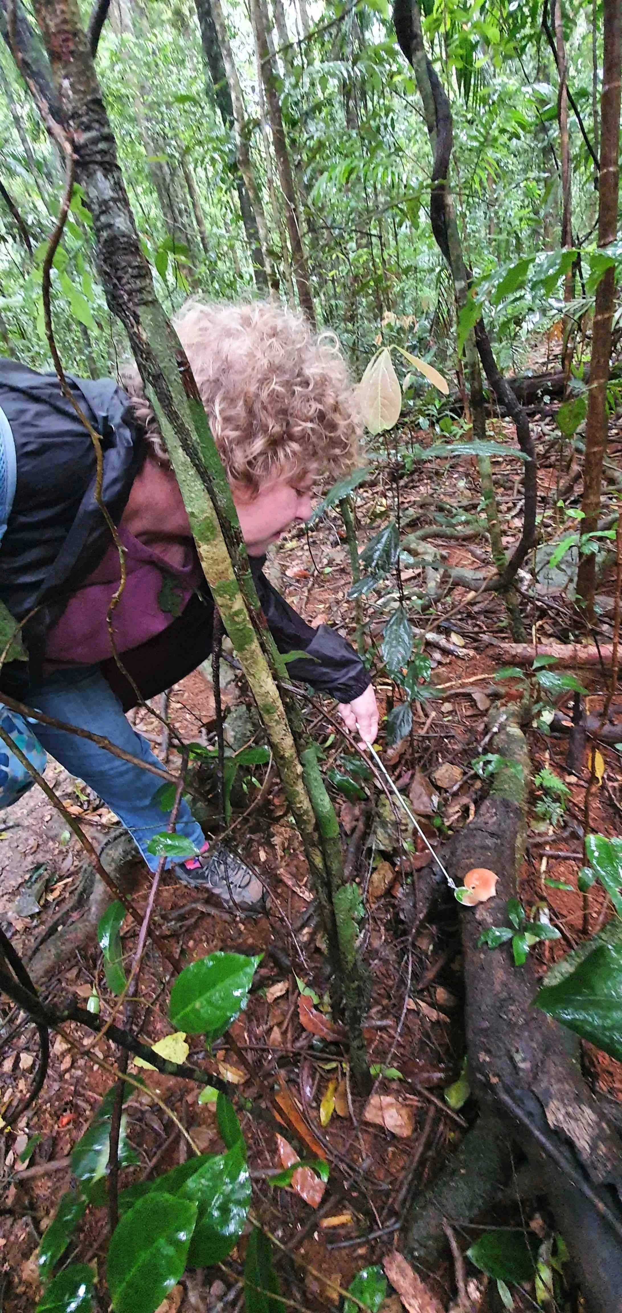
Orange Fan - Anthracophyllum archeri
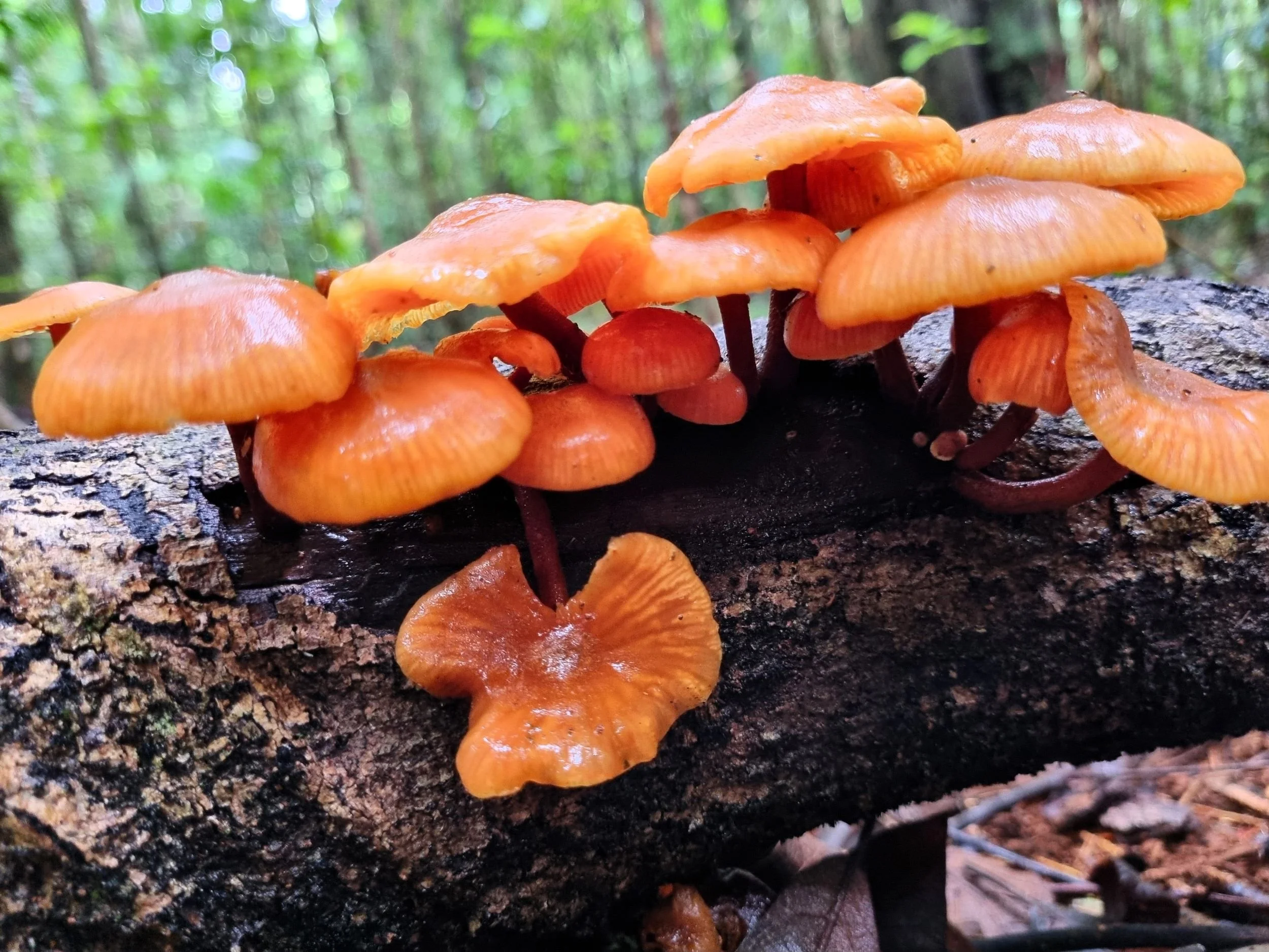
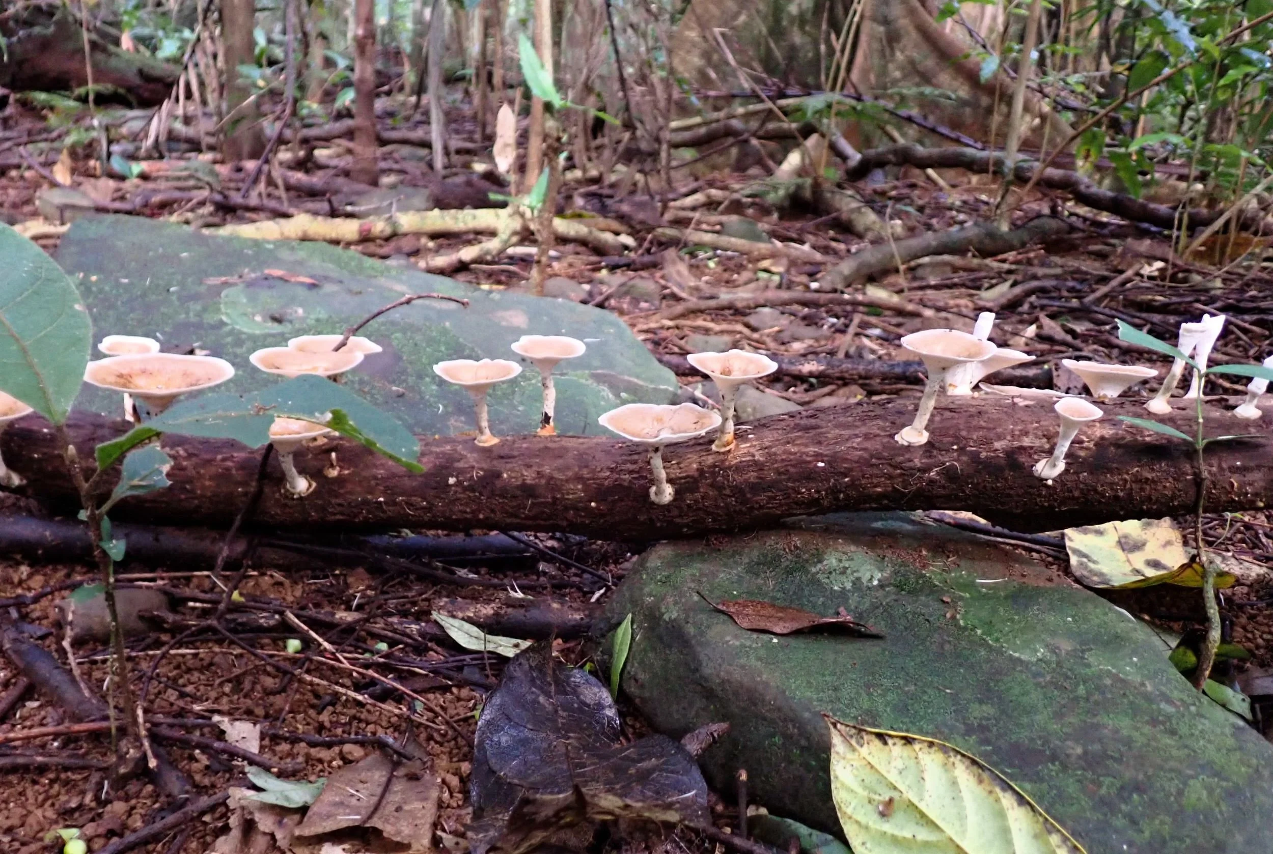
Orange Faint Foot Mushroom - Heimiomyces tenuipes

Gill pattern of the Orange Faint Foot

Group of Orange Faint Foots with scale


Leucocoprinus cretaceus (?)

Orange-scruffy Collybia - Cyptotrama asprata

More Orange-scruffys - earlier stage of development

Pluteus sp.?

Underside of ? Pluteus sp.


Coprinellus sp.

Heimiomyces sp.?


Yellow Flower-Pot - Leucocoprinus birnbaumii

Marasmius sp. 'pinwheels and parachutes mushrooms'

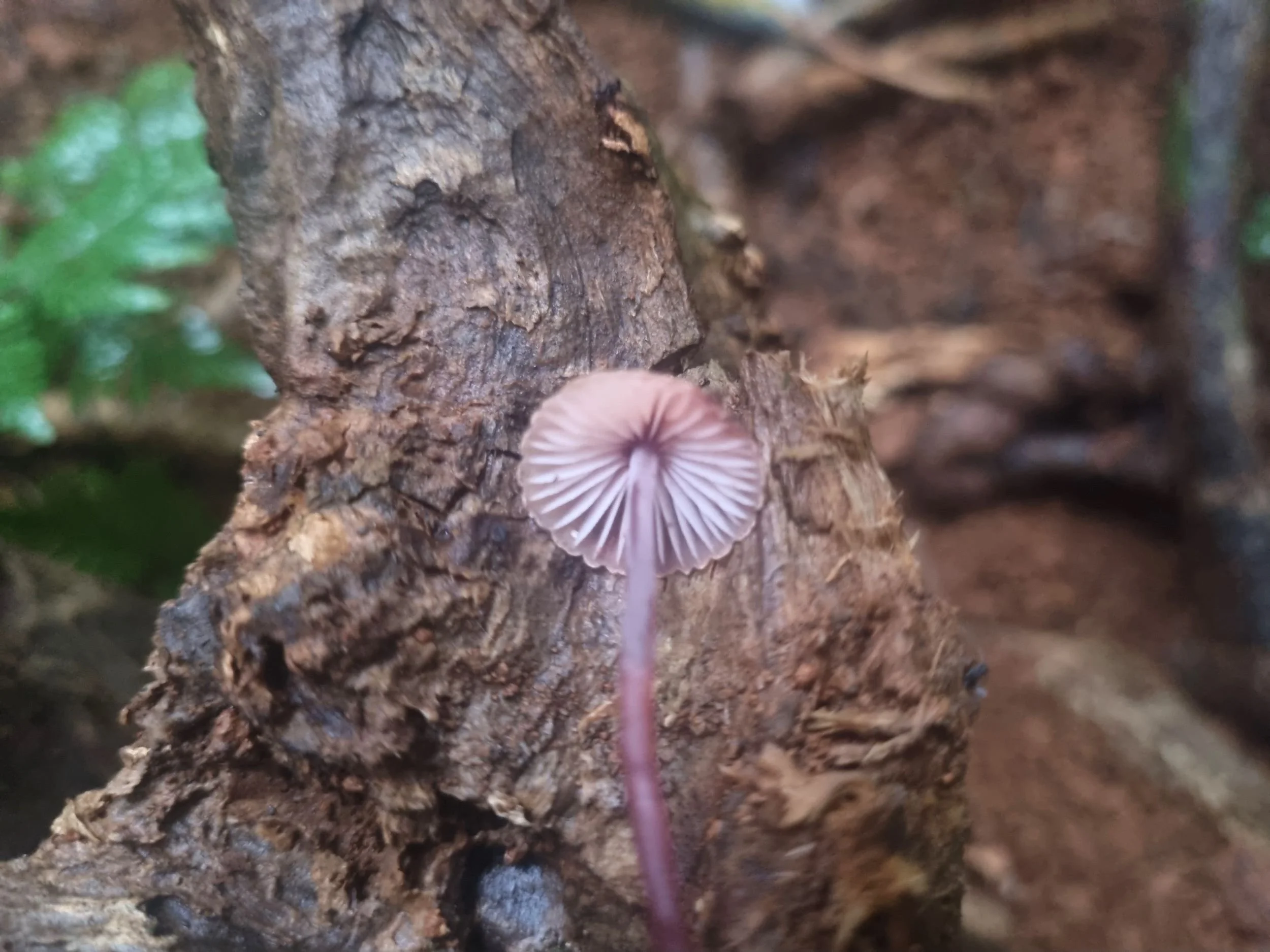
Ruby Bonnet - Cruentomycena viscidocruenta


Ruby Bonnet with scale

Troupe of Mycena sp.?

Porcelain Slimecap - Oudemansiella australis

Porcelain Slimecap

Orange Ping-Pong Bat - Favolaschia calocera

White Porecap - Favolaschia pustulosa

Top of Fav. pustulosa

Could be Omphalotus nidiformis - ghost fungus


Collybia sp.?


Mycena or Hygrocybe sp.?


Fan species - Campanella?

Australian shitake - Lentinula lateritia


Mycena kuurkacea

Mycena sp.

Gills of previous Mycena

Coprinellus diseminatus

Polypores, Leathers and Crust Fungi
Polypores have ‘pores’ not gills, and the pores can vary from quite large to microscopic so you need a hand lens to see them. They are often hard and woody or leathery textures with the fruiting body like a shelf or bracket that is attached directly to their substrate.
Leathers can look like polypores but their lower surfaces have no pores. They may have stems, or not, and sometimes form dense overlapping colonies. The distinctive brown and orange Stereum ostrea is a leathery shelf-fungi often seen in colonies along dead logs in Mary Cairncross.
Crust, paint or patch fungi adhere to the substrate, usually decaying wood, in a flattish layer like a splash of paint, or with some texture like a raised edge or a soft cushion. Most have spores but these may be microscopic.

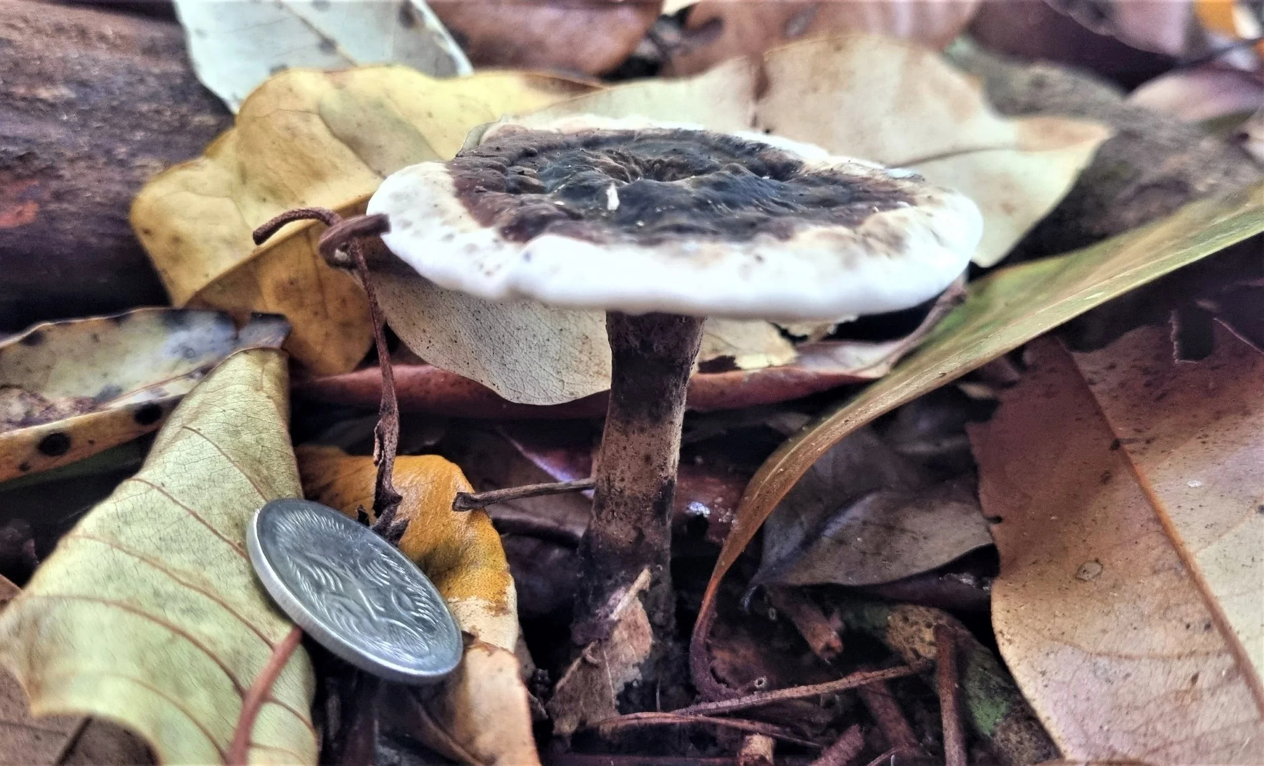
Checking underside of Sanguinaderma rude

Small scratch shows characteristic red staining of Sanguinaderma rude


Stalked polypore sp.


Fomitopsis sp.?

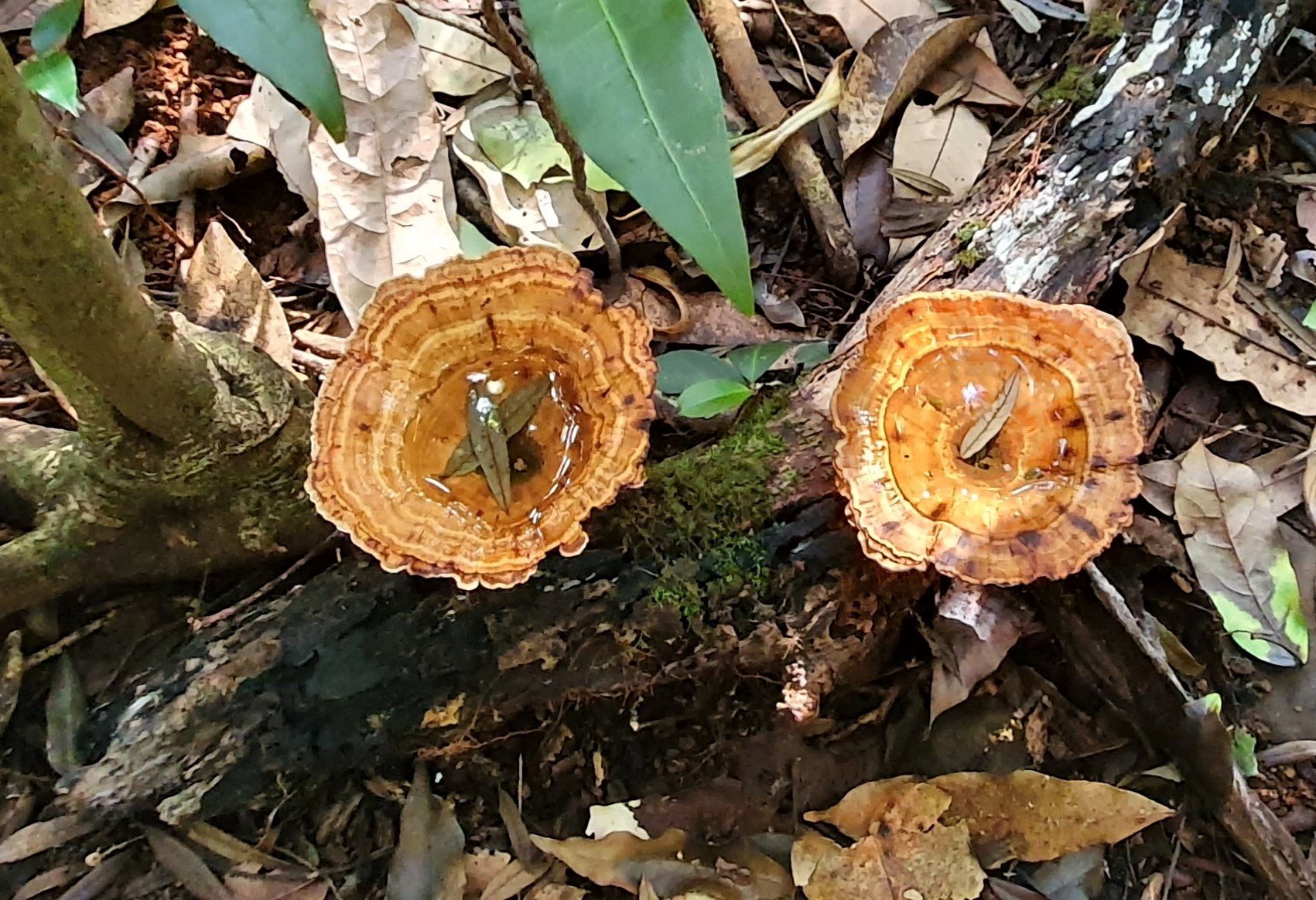
Microporus xanthopus - yellow footed tinypore

Microporus xanthopus

Microporus xanthopus

Ditto, showing characteristic funnel shape and 'yellow foot' (basal disc) attaching the stem to the substrate

Stereum ostrea

Stereum ostrea


Microporus affinis


Trametes versicolor
Jellies
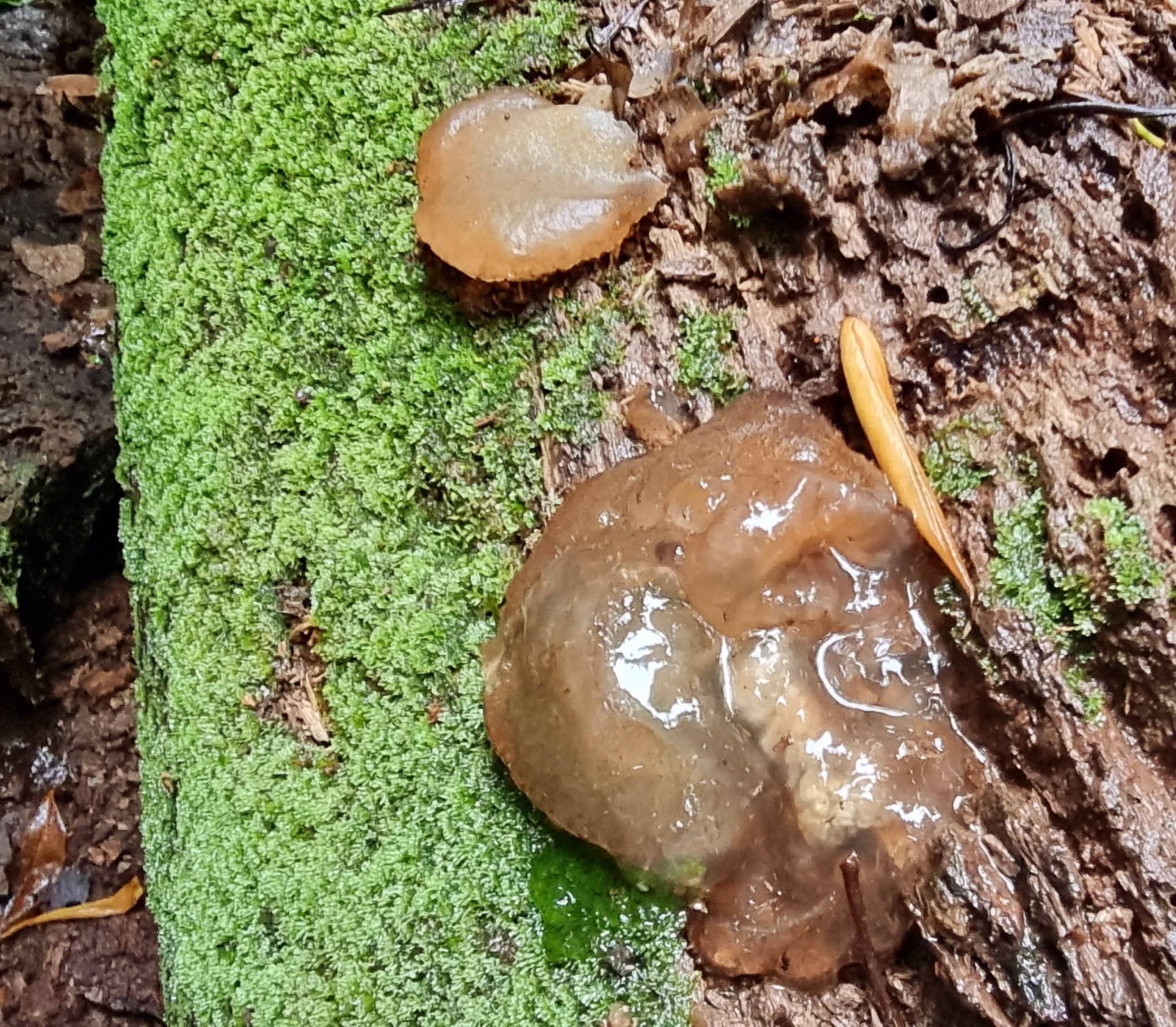
Jelly fungi have fruit bodies that are gelatinous in appearance and texture. They are usually found on wood or as parasites of other fungi. Frequently seen in Mary Cairncross are the Jelly Ear Fungus and Wood Ear Fungus, both are Auricularia species.

Auricularia sp.

Auricularia cornea?

Auricularia auricula-judae


Auricularia sp.


Tremella sp.

Dacryopinax spathularia

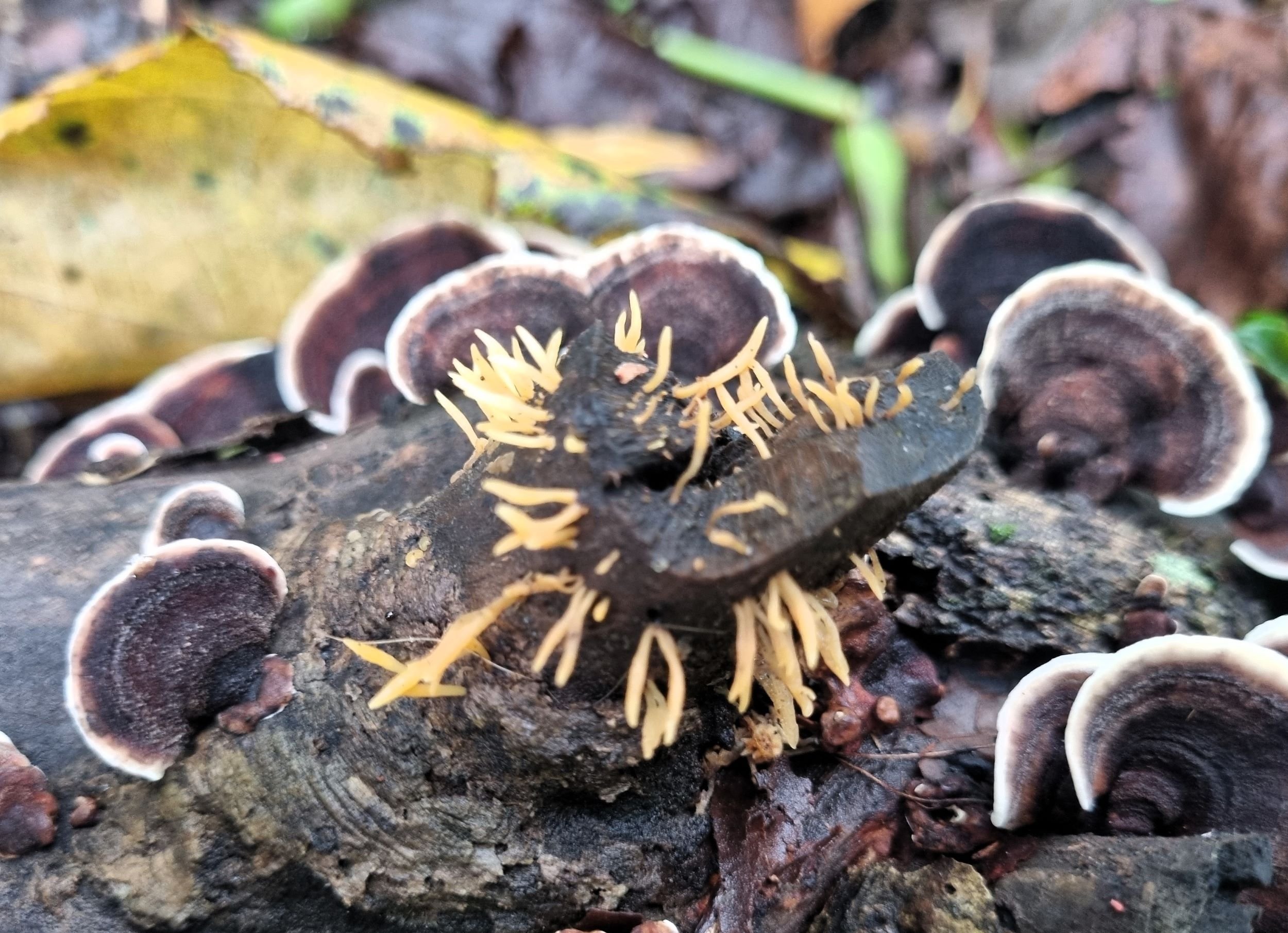
Calocera sp. amidst polypores
Ascomycetes
Ascomycetes are commonly known as sac fungi - they produce spores in microscopic sacs called asci that form the fertile layer. It’s a hugely diverse group and the fruiting bodies come in varied forms including cups, discs, flasks, pins and club-shaped.

Phillipsia subpurpurea


Xylaria sp.


Xylaria sp.

Xylaria sp..

Ascomycota? or a resupinate fungi?

Chlorociboria sp.

Species under investigation

Same as previous
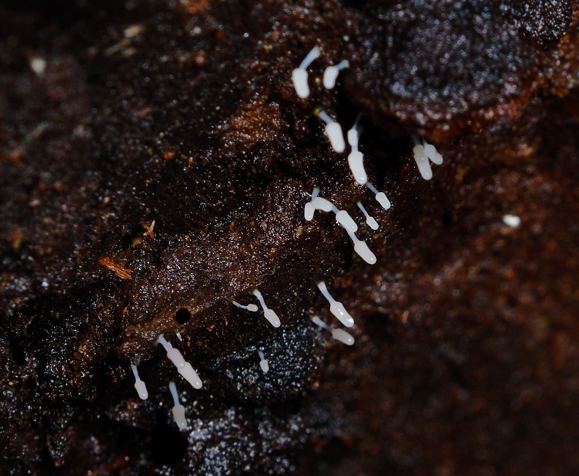
Slime Moulds - ‘The Myxomycetes’
Slime moulds are not true fungi - they are classified as Protista - neither plants, animals nor fungi. Plasmodial slime moulds, also known as myxomycetes, are at the reproductive life stage when they look superficially similar to fungi and reproduce via spores, and they can also move (observed via time-lapse as ‘pulses’ of movement as they feed on bacteria). They range in size from large amorphous blobs like the lovely ‘Dog Vomit’ slime mould to tiny delicate structures that require a hand lens to see and macro photography equipment to capture their features.

Lycogola sp.

Lycogola epidendrum - Wolf's Milk

Ceratiomyxa fruticulosa - Icicle Fairy Fans

Theresa checking out wood heap for slime molds

Stemonitis sp. - Chocolate Tube Slimes

Stemonitis sp.


Fuligo septica - dog vomit slime mould

Slime mold (yellow) in the plasmodial stage covering a Panellus sp. possibly luxfilamentus

Ditto, with scale included
Slime mould, query sp.
Puffballs, Stinkhorns, and Birds Nest Fungi
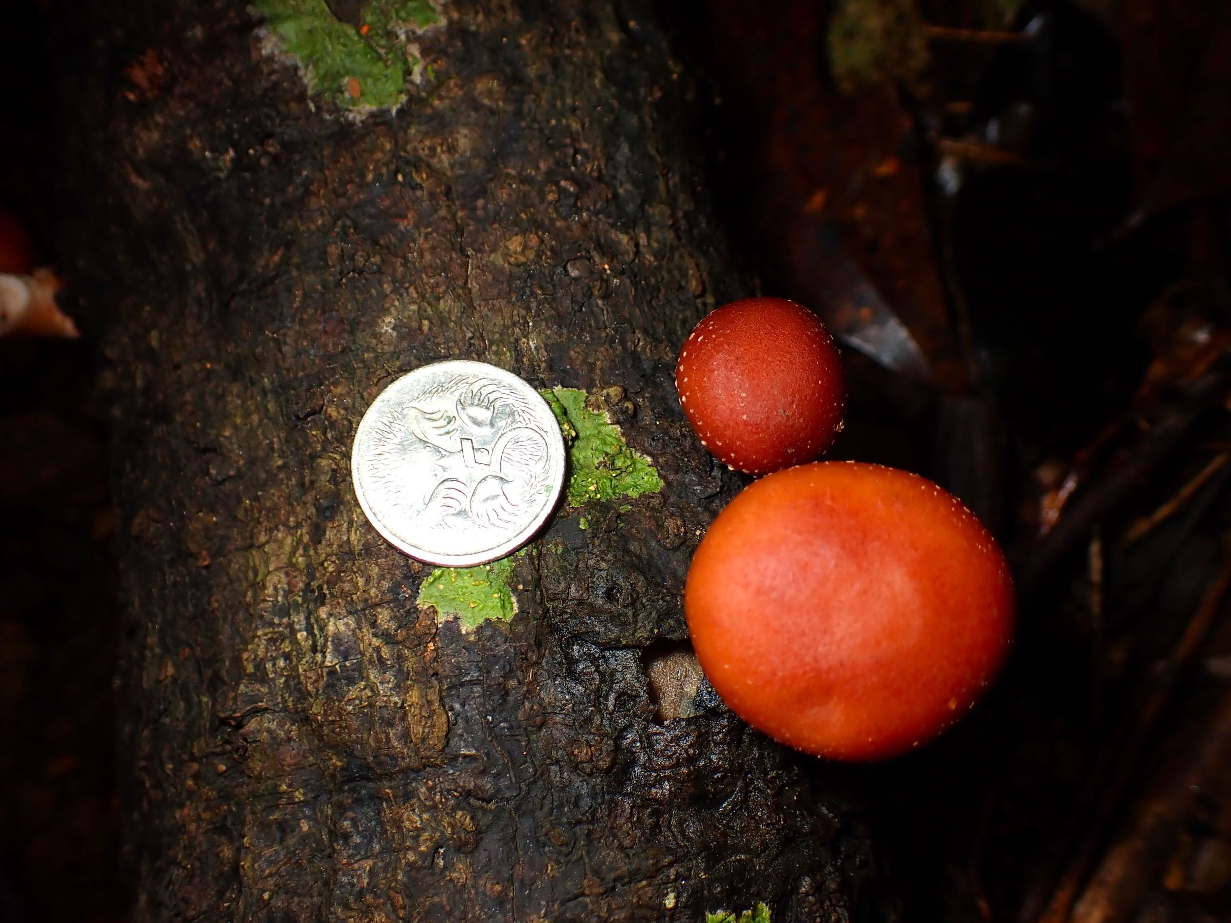
Puffballs are usually ball-shaped in form. They produce spores internally - as they mature the inside becomes filled with their dry and powdery spores which are actively released by the action of wind or rain or similar disturbance. The spores are released through an apical hole (on the top of the ball) or the whole outer skin breaks open. Earthstars are related - the ball-shaped spore case is surrounded at maturity by the star-like arms of the outer wall layer, which serve to elevate the spore case from its immediate surroundings facilitating spore dispersal by wind and rain.
Stinkhorns! These fabulous fungi with their weird forms and unpleasant stinky smell are favourites of ours. The fruiting bodies emerge from an egg-like sac as it rapidly expands. The spore mass is in a gelatinous ‘gleba’ emerging from the receptacle and typically smells of dead flesh, semen, or dung, and attracts flies, beetles and other insects to help disperse the spores.
Birds Nest Fungi are small, steep-sided cups (up to a centimetre across and of similar height) which contain small disk-like objects, giving the appearance of eggs in a nest. These eggs are periodoles or ‘spore packets’ and contain the spores. The spores are ejected when rainwater splashes into the cup.

Lycoperdon sp.


Lycoperdon sp. with opening

Lycoperdon sp. possibly subincarnatum

Lycoperdon sp. - close-up


Lycoperdon sp.

Geastrum sp. possibly triplex

Geastrum sp. possibly triplex

Aseroe rubra - Starfish stinkhorn

Phallus multicolor, newly emerged from sac

Egg sac and veil of Phallus multicolor

Veil and apical opening


Birds Nest fungi - found nearby Mary Cairncross
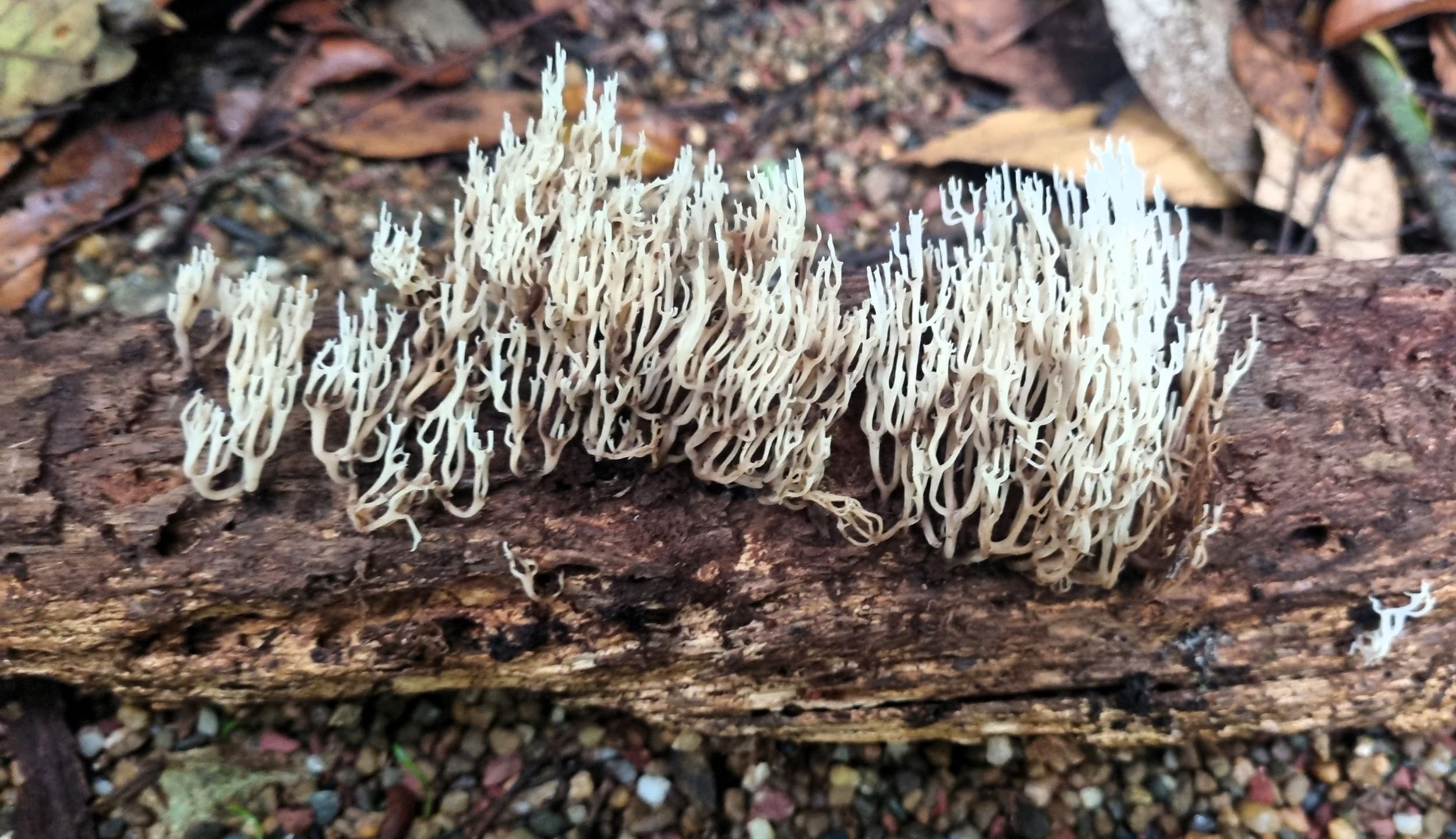
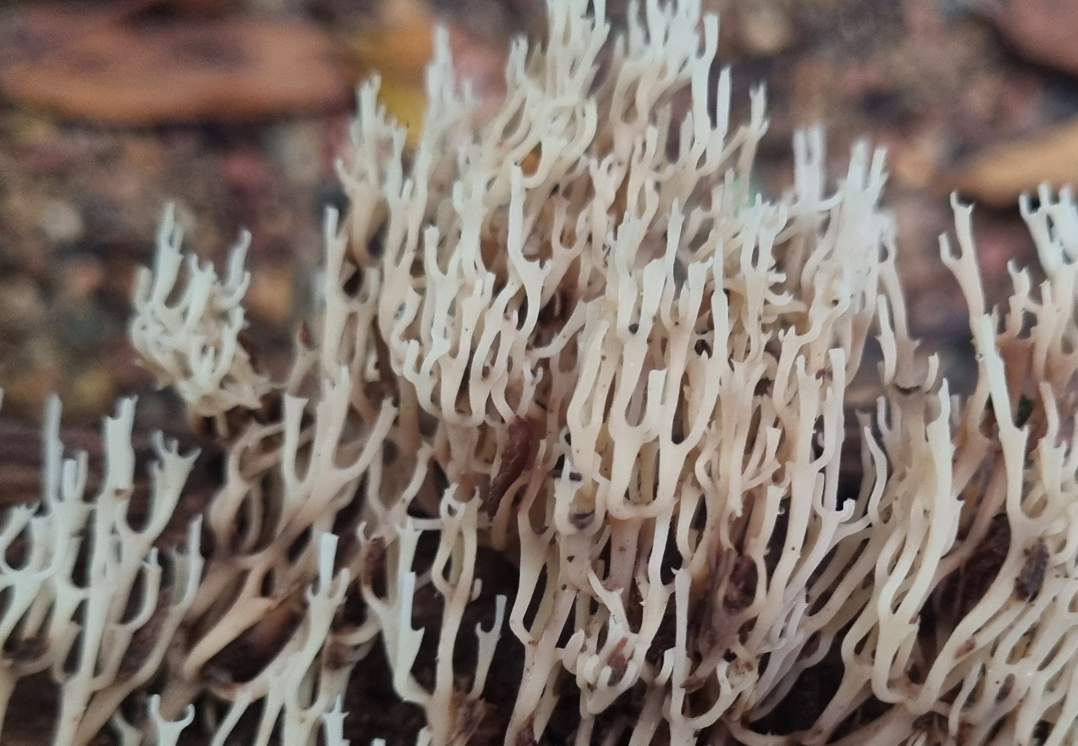
Corals
Coral fungi range from a simple unbranched club shape to large branching coral-like structures. Spores are produced all over the body except the stem.